Research - Early detection of Alzheimer’s using computer vision
Introduction

Illustration of the eye
Alzheimer’s Disease (AD) is an irreversible and progressive disease of the brain
Type 2 diabetes commonly leads to AD
Often detected at a late stage
Lack of cure
Leads to death
No reliable early detection methods
New research shows AD patients have less choroidal thickness and can be used as a biomarker for early detection of AD
Research Question
Can choroidal thickness be accurately measured?
Method

Figure shows OCT image of the cross-sectional view of the eye
Choroidal space is visible in Optical Coherence Tomography (OCT)
OCT scans are available publicly for diabetic retinopathy
No scans available for AD patients publicly
Develop algorithm to automatically identify the choroidal thickness
Test the algorithm on scans available for diabetic retinopathy
Identify the accuracy of the choroidal thickness
Testing
Downloaded OCT scans from public database
Selected 206 scan results for Normal and Diabetic Retinopathy
Sub-selected scans that are horizontal scan from nasal to temporal region
Total of 40 scans selected for testing
Downloaded Manual Segmentation data
Experts manually marked the retinal segmentation
Total of 25 scans were manually scored
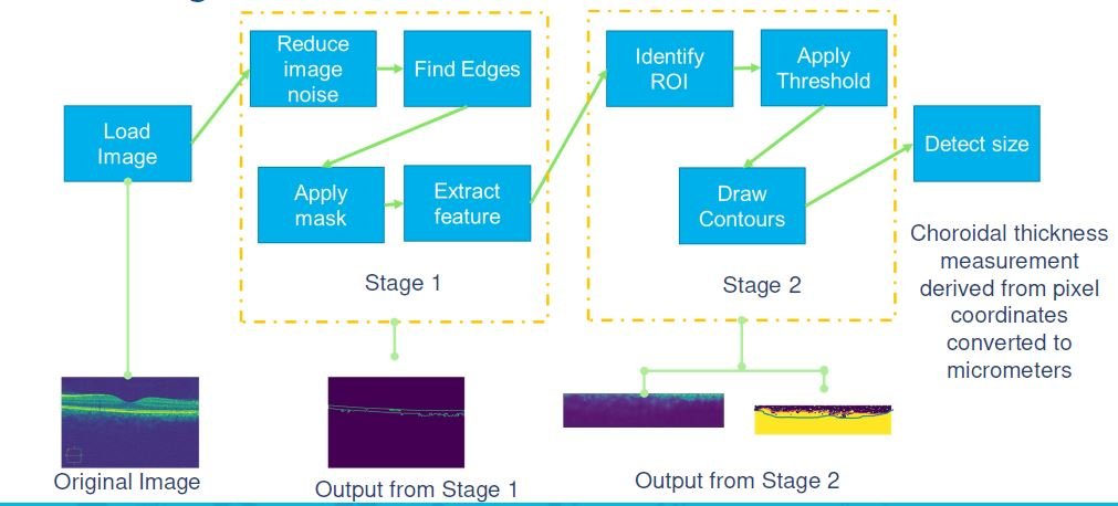
Developed novel software algorithm to measure the thickness automatically
Developed novel software algorithm to measure the thickness automatically
Executed the algorithm using Python 3.0 on Linux OS
Statistically compared the results from the algorithm with the manual scoring
Novel Algorithm
Results

40 different scan results were tested
20 Normal and 20 Diabetic Retinopathy
Best results could be obtained with black background and contrast enhancements
Variability in size was dependent on scan configuration
Size variability was minimal when the scan output was flat (Error < 0.02% p value = 0.001)
Size variability was significant when the scan was done at an angle (Error > 1% p value <0.005)
The sizes detected using the algorithm are found to be matching the ground truth measurements
Overall Error rate: ±0.02% (p value = 0.001)
Discussion
Several publicly available databases have OCTB scans
Available for normal eye, diabetic retinopathy and other retinal diseases
No scans available for Alzheimer’s patients
Used of diabetic retinopathy for the test
Built a novel algorithm to use the geometry of the scans to find the thickness
Results were measured against the manual segmentation
Error rates were less than 0.02%
Less than 4 micrometers difference
References
Gholami, Peyman, and Lakshminarayanan, Vasudevan. Optical Coherence Tomography Image Retinal Database. Ann Arbor, MI: Inter-university Consortium for Political and Social Research [distributor], 2019-02-20. https://doi.org/10.3886/E108503V1
Patton N, Aslam T, Macgillivray T, Pattie A, Deary IJ, Dhillon B. Retinal vascular image analysis as a potential screening tool for cerebrovascular disease: a rationale based on homology between cerebral and retinal microvasculature J Anat. 2005; 206:319–348. [PubMed: 15817102]
De Silva DA, Manzano JJ, Woon FP, Liu EY, Lee MP, Gan HY, Chen CP, Chang HM, Mitchell P, Wang JJ, Lindley RI, Wong TY, Wong MC. Associations of retinal microvascular signs and intracranial large artery disease. Stroke. 2011; 42:812–814. [PubMed: 21257821]
Ermengarda Marziani; Simone Pomati; Paola Ramolfo; Mario Cigada; Andrea Giani; Claudio Mariani; Giovanni Staurenghi, Evaluation of Retinal Nerve Fiber Layer and Ganglion Cell Layer Thickness in Alzheimer's Disease Using Spectral-Domain Optical Coherence Tomography
Fang He ,1 Rachel Ka Man Chun ,2 Zicheng Qiu ,1 Shijie Yu ,1 Yun Shi,3 Chi Ho To, Choroid Segmentation of Retinal OCT Images Based on CNN Classifier and - Fitter